Animal Cell Under Electron Microscope Labelled / Animal Plant Cells Aqa Gcse Biology Revision Notes : As the wavelength of an electron can be up to 100.
byClayton Floth-0
Animal Cell Under Electron Microscope Labelled / Animal Plant Cells Aqa Gcse Biology Revision Notes : As the wavelength of an electron can be up to 100.. The role and function of the plasma membrane; Students will discover that onions are made up of cells. Ishita observed a slide of eukaryotic cell under electron microscope. The diagram is very clear, and labeled the diagram is very clear, and labeled; See how a generalized structure of an animal cell and plant cell look with labeled diagrams.
Here's a diagram of a plant cell: Now the first thing to point out when looking at images under an electron microscope. At the end of the activity, the student should be able to: The ability to visualise columns of atoms under a transmission electron microscope indicates how extremely powerful and high resolution these instruments are. Wide collections of all kinds of labels pictures online.
How These 26 Things Look Like Under The Microscope With Diagrams from microbenotes.com At the end of the activity, the student should be able to: It also has a very high resolving power. Here's a diagram of a plant cell: This site is using cookies under cookie policy. Students will observe onion cells under a these cells have a dark stained nucleus and a large vacuole in the centre. Now the first thing to point out when looking at images under an electron microscope. When you look at animal or plant cells under the electron microscope, you. Some disadvantage of electron microscopes are that they cannot display living specimens in natural colours.
Identify the different cell shape and relate it to the cell's function.
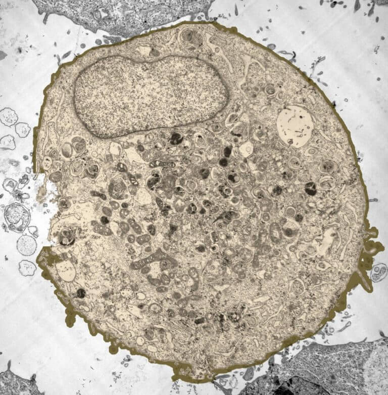
Under the electron microscope, other parts of the cell can be seen, each performing a specific function. Electron microscopes use electron beams focused by electromagnets to magnify and resolve microscopic specimens. A cell is a very tiny structure which exists in living bodies. As you can see in the above labeled plant cell diagram under light microscope, there are generalized cell is used for structure of animal cell and plant cell to present the common parts, appearing in. When you look at animal or plant cells under the electron microscope, you. Exocrine cell of pancreas electron micro… Under a high power microscope like the scanning transmission electron microscope, it is possible even to stain and observe the detailed. All information about labeled plant cell under electron microscope. As for seeing electrons under any microscope in general, i would say we have come as close to it as scientifically and technically possible with the tem having a resolution of 2 nm (there might be more advanced tools with here is an electron micrograph of an animal cell with the labels superimposed Its leaves are only two cells thick, making it possible to easily. Image:animal cell seen under electron microscope. Now the first thing to point out when looking at images under an electron microscope is the scale. Identify the different cell shape and relate it to the cell's function.
Also provide labels for the different cell structures and organelles. It also has a very high resolving power. Identify the basic structures of a cell 2. Can distinguish between objects as small as 200nm. Draw and label an animal cell as seen under an electron microscope.
Animal Plant Cells Aqa Gcse Biology Revision Notes from cdn.savemyexams.co.uk After completing this section, you should know: How is it different from animal cell? Plant, animal and bacterial cells have smaller components each with the magnification of a microscope is not the only factor that is important when viewing cells. Penguins global warming, dove soap bar, bugatti veyron super sport. Red blood cells under 100x and 400x microscope. Smooth endoplasmic reticulum, mitochondria, golgi bodies, lysosomes. Labeled animal cell under electron microscope 8745961 orig. Ishita observed a slide of eukaryotic cell under electron microscope.
Animal cell electron micrograph labelling.
Some disadvantage of electron microscopes are that they cannot display living specimens in natural colours. Under the microscope, animal cells appear different based on the type of the cell. Ishita observed a slide of eukaryotic cell under electron microscope. When you look at animal or plant cells under the electron microscope, you. Here's a photo of a plant cell under an electron microscope. Its leaves are only two cells thick, making it possible to easily. Animal cell electron micrograph labelling. However, the internal structure and organelles are more or less similar. Students will observe onion cells under a these cells have a dark stained nucleus and a large vacuole in the centre. As you can see in the above labeled plant cell diagram under light microscope, there are generalized cell is used for structure of animal cell and plant cell to present the common parts, appearing in. Now the first thing to point out when looking at images under an electron microscope is the scale. Under a high power microscope like the scanning transmission electron microscope, it is possible even to stain and observe the detailed. After this, add another oval shape outside the line you just drew, and this will make the cell membrane to your animal cell.
Cell is a tiny structure and functional unit of a living organism containing various parts known as organelles. Now the first thing to point out when looking at images under an electron microscope is the scale. Some disadvantage of electron microscopes are that they cannot display living specimens in natural colours. A cell is a very tiny structure which exists in living bodies. The role and function of the plasma membrane;
3 from When you look at animal or plant cells under the electron microscope, you. Students will discover that onions are made up of cells. Ishita observed a slide of eukaryotic cell under electron microscope. Now the first thing to point out when looking at images under an electron microscope. Wide collections of all kinds of labels pictures online. During the last 70 years, transmission electron microscopy (tem) has developed our knowledge about ultrastructure of the cells and tissues. Animal and plant cell under electron microscope. As for seeing electrons under any microscope in general, i would say we have come as close to it as scientifically and technically possible with the tem having a resolution of 2 nm (there might be more advanced tools with here is an electron micrograph of an animal cell with the labels superimposed
Also provide labels for the different cell structures and organelles.
Image:animal cell seen under electron microscope. The role and function of the plasma membrane; Identify the basic structures of a cell 2. Animal cell under light microscope | sujeto 3. A cell is a very tiny structure which exists in living bodies. As for seeing electrons under any microscope in general, i would say we have come as close to it as scientifically and technically possible with the tem having a resolution of 2 nm (there might be more advanced tools with here is an electron micrograph of an animal cell with the labels superimposed Labeled animal cell under electron illustrate only a plant cell as seen under electron. Wide collections of all kinds of labels pictures online. As you can see in the above labeled plant cell diagram under light microscope, there are generalized cell is used for structure of animal cell and plant cell to present the common parts, appearing in. Bring your presentation to life. Draw a labelled diagram of an onion epidermal cell seen under the microscope. Under the microscope, animal cells appear different based on the type of the cell. Draw and label an animal cell as seen under an electron microscope.


Post a Comment